Research topic
(Back)
X-Ray imaging
Pathologies in the vascular system (angiopathies), and in particular those concerning the heart (cardiopathies)
are the first death cause in Europe. They are mostly due to stenosis formations: lipids deposits reduce or occlude the vessels.
The consequences can be very serious especially when they impact arteries that are as crucial to the organisms life as coronaries
(which provide the heart in oxygen).
X-Ray imaging offers a minimally invasive mean to diagnostic and to treat such malformations. The clinician introduces through
the patient's groin the interventional tools he needs, and handles them remotely, guiding his gesture based on the images acquired
with X-Rays.
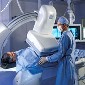 General Electric Innova imaging system
 |
|
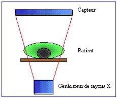 X-Ray image formation
 |
Difficult interventional images
The X-Ray imaging system can be used both for diagnostic or interventional purposes. In the first case relatively high radiation
are allowed, resulting in well contrasted images. On the other hand, interventional exams are carried out under limited radiation and
must thus be digitally denoised.
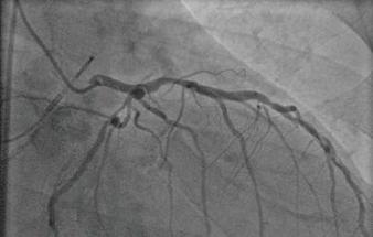 Typical image for diagnostic exams
 |
|
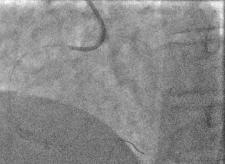 Typical image for difficult fluoroscopic exams
 |
Since spatio-temporal denoising techniques present difficult problems of noise coloration, our work focuses on pure temporal
filtering only. My PhD aims at overcoming the main limitation of temporal filters: its unability to handle motions.
(More)
Transparent images
X-Rays are attenuated in various ways according to the materials they are going through: different organs react in different
ways to the same radiation. It is thus possible to visualize the internal anatomy of a patient, by exposing him to a X-Ray radiation
and by counting locally the X photons coming through.
But two organs set over each other attenuate the radiation successively: their attenuations multiply themselves. There is no
occlusion in the world of radiations but multiplicative transparencies.
Moreover, since the measured images are easier to interprate in the world of physical thicknesses than in the world of radiations,
a log()-operator is applied to them which transforms the multiplicative in additive transparency.
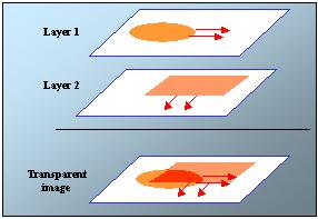 Layer model
 |
|
 Transparent fluoroscopic image
 |
For instance, we can see on the example above the heart superimposed to the diaphragm, the lungs, the spine, the ribs and the interventional
devices.
Thus, because of the specificity of X-Ray images, the image formation model is totally different from the classical video images
model. In particular, the brightness consistency over the objects' trajectory is not verified. Since most of motion estimation methods
are based on this assumption, new frameworks have to be specifically designed for transparent motions.(Back)
My PhD thus aims to answer two questions to solve our problem:
How can we estimate the motions present in the image?
How can we use that information in an efficient temporal denoising?
Our contributions on these two topics are briefly introduced in the next page.
Back